KS3 Biology › Cells

KS3 Biology › Cells
These are the points the worksheet questions are built around.
Cells might look flat in textbook diagrams, but real cells aren't flat at all — they're three-dimensional, with length, width and depth, just like a real ball looks completely different from a flat circle drawn on paper. In 3D, an animal cell looks more like a blob or bubble with structures floating inside it. Cells come in a huge range of shapes and sizes depending on their job: red blood cells are shaped like squished frisbees, nerve cells are long and thin, and muscle cells are stretched-out tubes — all still fundamentally 3D, just specialised for what they do.
Cells are far too small to see with the naked eye — around 100 of them could fit across the width of a single full stop. That's why scientists use light microscopes to magnify them. Even then, some structures inside a cell are still too small to see clearly, which is why even more powerful electron microscopes are sometimes needed.
Every animal cell shares four main structures. The cell membrane is a thin protective layer surrounding the whole cell, controlling which substances can enter (like nutrients and oxygen) and which can leave (like waste products) — without it, the cell would simply fall apart. The cytoplasm is the gel-like substance filling most of the cell, acting as its workspace: this is where chemical reactions happen, including building proteins, breaking down nutrients, and releasing energy.
The nucleus acts as the cell's control centre. It contains DNA — the genetic information that tells the cell what to do, including instructions for making proteins and carrying out its everyday activities. Mitochondria are sausage-shaped structures scattered through the cytoplasm, often called the powerhouses of the cell, because their job is to release energy through cellular respiration, using oxygen and food.
Every animal — whether human, cat, beetle, jellyfish or elephant — is made of cells containing these same four structures. Some cells become specialised for particular jobs: red blood cells carry oxygen, nerve cells send electrical signals (and can be over a metre long, to reach from the spinal cord to the toes), muscle cells contract to create movement (and are packed with extra mitochondria, since movement needs a lot of energy), and sperm cells swim to fertilise eggs. Underneath all these specialisations, the same basic structures are still there.
Because cells are so difficult to see and understand directly, scientists represent them using models — simpler versions of something complex, similar to how a toy car represents a real car in an easier-to-understand way. Models can be 2D, like a flat textbook diagram, or 3D, built from everyday materials — a ball for the nucleus, pasta for the mitochondria, a jelly-filled bag for the cytoplasm.
Not every model is equally accurate, so scientists evaluate them — identifying what a model shows well and what could be improved. For example, a plastic bag might represent a cell membrane reasonably well, since it surrounds everything the way a real membrane does. But a real cell membrane has tiny gaps that let specific substances through, which a plain bag doesn't do — so poking small holes in it, or swapping it for holed cling film, would be a genuine improvement.
In short: animal cells are three-dimensional, and every one shares four key structures — the cell membrane, cytoplasm, nucleus and mitochondria — each with a distinct job. Cells become specialised for different roles while keeping these same basic structures, and because cells are so small and complex, scientists use 2D and 3D models to understand them, evaluating each model's strengths and weaknesses along the way.
These are the words that appear as key term cards in the video — worth knowing cold before the test.
Short clips from this lesson. Good for a quick refresh before a test.
Swipe sideways to see more →
Revising for a test? Key terms, objectives and quiz questions on one printable page.
The 3 questions from the video, one at a time. See if you caught them.
1. Which of these best describes the shape of real animal cells?
Why: Real cells are three-dimensional, like tiny blobs or bubbles. Textbook drawings are flat because that is easier to draw.
2. Roughly how many animal cells could fit across the width of a single full stop?
Why: A full stop looks tiny, but animal cells are much smaller still. About 100 of them would fit across it.
3. Why are muscle cells packed with extra mitochondria?
Why: Muscles contract to make you move, which takes a lot of energy. Mitochondria release energy from food, so muscle cells need lots of them.
Score: 0 out of 3 —
Want more practice? Try the 60-second Quiz Challenge →
This lesson is a level in our game Keep the Cell Alive
One page with a full answer key. Enough to check the lesson landed, and to see how the questions are written before you buy anything.
Get the free worksheet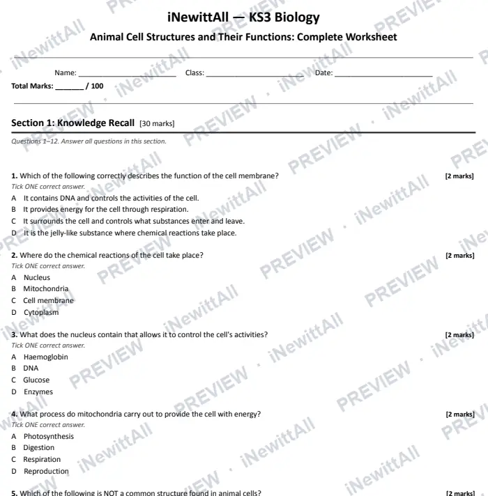
The top of page 1. The answer key is included when you buy.
£5 £6
The full worksheet and the extension pack together, with both answer keys.
Instant download with full answer keys. Print as many copies as your class needs.
£3
£3
£5 £6
Tick it off to track your progress. It's saved on this device. See my revision →
Students watch the video and do the quiz. They get a result card to paste back to you, showing their first try and best score.
This lesson's quiz questions on one sheet, with the answer key on page 2.
The quiz in big text for your classroom screen, one question at a time. Or build a 5-question Do Now starter from lessons your class has already done.
You're welcome to embed the video and link these resources in Google Classroom, Stile, Canvas, Compass or any other school platform. No need to ask.
Everything here is written to the KS3 curriculum for students aged 11–14. Print freely for the classes you teach.
 Next lessonPlant Cell Structures and Their Functions
More in this unit: Cells
No spam, unsubscribe anytime. You must be 13 or over to sign up.